
The ability to visualize inside the body has improved dramatically since Dr. Wilhelm Roentgen discovered the x-ray in 1895. Although a revolutionary concept at that time, soon after that discovery, doctors began specializing in using x-rays to diagnose disease. Since then, numerous discoveries resulting in more sophisticated techniques have improved the examination process.
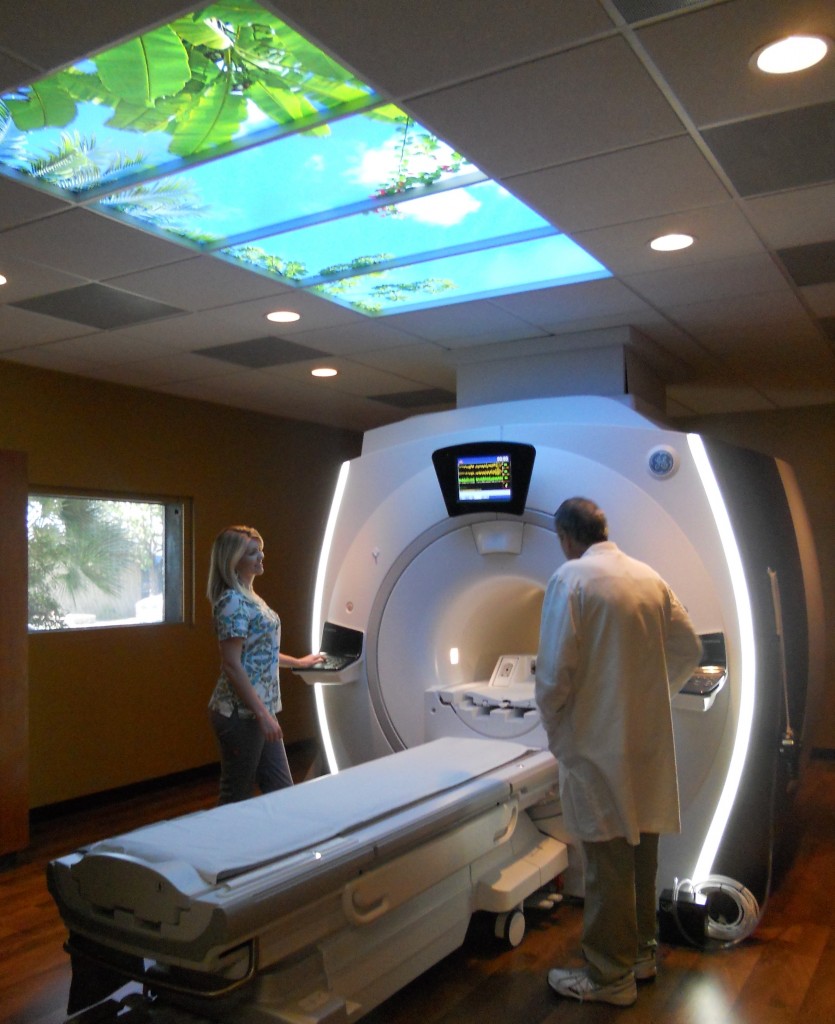 Visalia is the home of the latest in this technology, the 3.0 Tesla Magnetic Resonance Imaging (MRI) machine, which sets a new standard by producing a high quality image. This technology provides a valuable service to the county with critical diagnostic studies now done locally, saving patients travel time and effort.
MRI helps doctors determine if a patient needs surgery, and is so, what kind of surgery. An MRI is often the final factor for moving treatment forward and it offers other benefits to the patient. There is no ionizing radiation exposure to the patient, and the faster scanning time, enlarged bore (opening) and cushioned table provide for a more comfortable experience.
MRI uses radio waves and a very powerful magnetic field to selectively excite slices of tissue within the body. As the tissue releases this energy as radio waves, a computer generates images.
“This magnet is similar in strength to the one they use to pick up cars at the junkyard,” said Glade Roper, MD, who completed a fellowship in musculoskeletal radiology at the Mayo Clinic in Scottsdale, AZ. “The more powerful the magnet, the higher the quality the image will be.”
Due to the exceptional anatomic detail, Roper noted the increased image clarity is particularly beneficial for medical conditions involving the brain, spine and musculoskeletal systems. Additionally, the greater contrast better differentiates between ligaments and tendons.
“It’s like watching baseball on an HD TV versus a traditional TV,” explained Roper. “On a regular TV, you can see the baseball leave the pitcher’s hand. In HD, you can see the stitches on the ball.”
“I am able to more accurately report pathologic conditions such as symptomatic herniated discs and potentially lead to earlier detection of disease processes affecting many people here in the Valley with diagnoses such as Alzheimer’s and Parkinson’s Disease,” said Dr. Aaron Berkey, a UCSD fellow trained in neuroradiology.
The 3T MRI is located at Visalia Imaging, 1700 S. Court St., Visalia. For more information, call 734-5674 or visit www.visaliaopenmri.com.